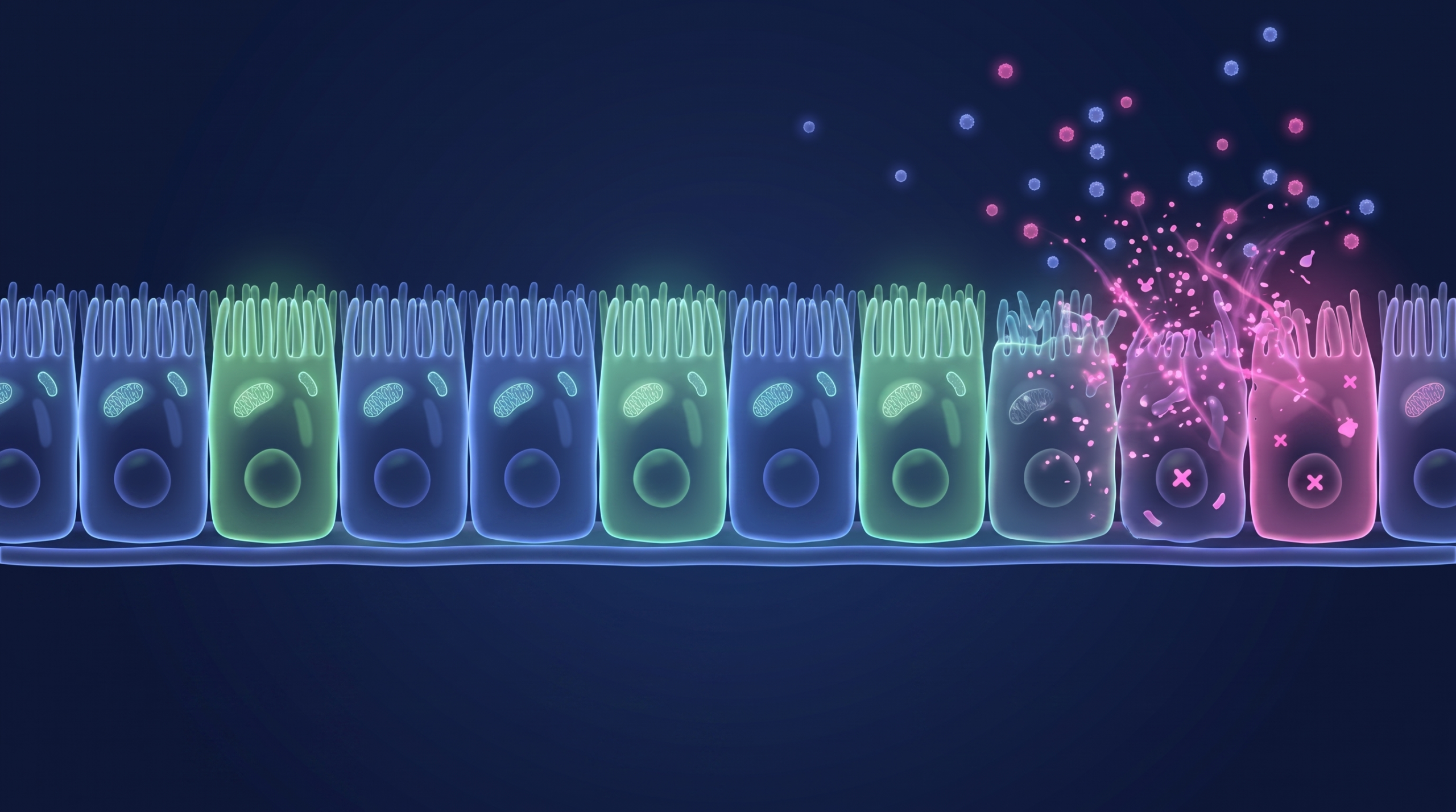
Ask most people to model gut inflammation and they reach for the same shortcut: pick a cytokine, dose it onto a monolayer, watch the barrier fall apart. The instinct is fine. The blind spot is treating the choice of cytokine as a detail. The two that matter most in inflammatory bowel disease, TNF-alpha and IFN-gamma, do not break the epithelium the same way. They run through different receptors, act on different clocks, and produce intestinal barrier dysfunction by mechanisms that barely overlap.
That is not an argument for always dosing them together. Plenty of programs study one pathway at a time, and should, because that is how you learn what a single mechanism actually does. It is an argument for a model that reproduces each one faithfully. If your compound acts on a pathway, that pathway is what you need to read correctly, and the model either recapitulates its biology or it does not.
The barrier is not incidental to IBD. It is close to the center of it, and the two cytokines reach it by almost opposite routes. Here is what each one does to the cells that face the lumen.
Different drugs act on different pathways
The distinction is not academic, because the IBD pipeline does not target “inflammation.” It targets specific nodes. The anti-TNF biologics, infliximab and adalimumab, act on TNF-alpha directly. JAK inhibitors like upadacitinib work further down, dampening the JAK-STAT signaling that IFN-gamma and other cytokines run through. Those are different mechanisms reaching different parts of the same inflamed tissue. Upstream of both, a switch like TL1A can release TNF-alpha and IFN-gamma together, which is part of why being able to separate their downstream effects is worth doing.
So the pathway you care about depends on what your compound touches. Work on a TNF-directed mechanism and TNF-alpha biology is the experiment. Work on the JAK-STAT axis and the IFN-gamma arm is. A model that blurs the two into a generic inflammatory response cannot answer either question cleanly.
TNF-alpha pulls the junctions open
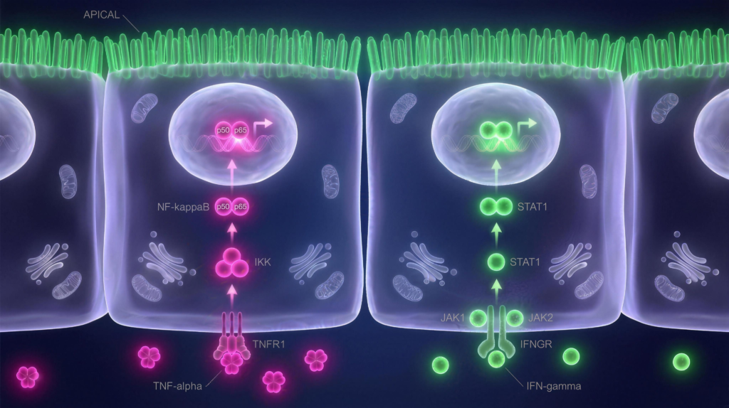
TNF-alpha signals through TNFR1 and, downstream, through NF-kB. That much is textbook. What matters for the barrier is where the signal lands. TNF-alpha drives expression of myosin light chain kinase, MLCK, and MLCK phosphorylates the myosin that rings each epithelial cell. When that actomyosin ring contracts, it physically tugs the tight junctions apart, widening the paracellular space between neighboring cells. The barrier does not dissolve. It gets pulled open from the inside.
The NF-kB step is not decoration. Work in intestinal epithelial monolayers showed that TNF-alpha’s increase in tight junction permeability requires NF-kB activation and the MLCK expression that follows it. Block the pathway and the contractile leak does not happen.
TNF-alpha has a second, blunter move. It drives apoptosis and cell shedding, and every shed cell leaves a transient gap in the sheet. In a three-dimensional model comparing the two cytokines directly, roughly 75 percent of TNF-treated epithelial structures contained apoptotic cells, against under 20 percent for controls and for IFN-gamma. Inhibiting caspases prevented the TNF permeability increase. It did nothing to the IFN-gamma effect. Two cytokines, two switchboards.
IFN-gamma strips the junction for parts
IFN-gamma signals through JAK-STAT1, and it works on a slower timescale. Its signature move is not contraction. It is removal. IFN-gamma drives the epithelium to internalize its own tight junction proteins, occludin, claudin-1, and JAM-A, through a macropinocytosis-like process, pulling them off the membrane and into the cell. Later work traced the route more precisely: a myosin II-dependent vacuolarization of the apical membrane that swallows the junction proteins into an internal compartment. The barrier fails because the bricks that build the junction have been carried inside.
IFN-gamma also primes the tissue, raising the epithelium’s sensitivity to TNF-alpha, in part by upregulating the TNF receptor itself, so a barrier already exposed to IFN-gamma responds harder to a later dose of TNF. In inflamed tissue the two often appear together. That interaction is real biology, and a faithful model should let you see it when you want to. It is not a reason to avoid studying either cytokine on its own.
Reading intestinal barrier dysfunction one pathway at a time
Because the two mechanisms are distinct, a system that only approximates “inflammation” is not good enough for pathway work. You need one where TNF-alpha produces its contractile, apoptotic signature and IFN-gamma produces its slower, endocytic one, each faithfully, so that when you dose a single cytokine the readout reflects that pathway’s real human biology rather than an averaged smear of both.
A model is only worth running if it reproduces the mechanism you are actually asking about. Dose TNF-alpha alone and you should see the junctions pulled open and cells shed. Dose IFN-gamma alone and you should see occludin and claudin carried inside without the apoptotic wave. The two can also act together, and in inflamed tissue they often do, synergizing through MLCK when both are present. A model that recapitulates each on its own terms lets you run any of those experiments, single agent or combined, and trust the result.
Turning “the barrier is breaking” into a number
Whichever pathway you dose, the damage converges on one thing you can measure: the barrier stops holding. The cleanest way to watch that happen is transepithelial electrical resistance, TEER. A healthy epithelial sheet is electrically tight, because ions cannot slip between the cells, so the layer holds a high resistance. When TNF-alpha’s contraction pulls the junctions apart, or IFN-gamma carries occludin inside, the paracellular path opens, ions leak through, and resistance drops. TEER turns barrier integrity into a single number, read in real time, without touching the cells. That is why it is the standard readout for barrier work.
It is also how the tissue reports back in practice. In InflammaScreen, TEER is measured at 24 and 48 hours after the inflammatory insult, next to an LDH cytotoxicity readout and IL-8 release, so a compound’s effect on the barrier, on cell survival, and on the inflammatory signal all come off the same monolayer. A dye tracer like Lucifer Yellow measures the same paracellular leak a different way, as an endpoint permeability flux rather than a live signal. For a barrier under cytokine attack, TEER is the more direct read, because it catches the junction opening as it happens.
How a human intestinal model interrogates this
This is the biology RepliGut® is built to reproduce, and it is where the choice of model stops being academic. Caco-2, the field’s default, is a colorectal cancer line. Its innate signaling is rewired, its tight junctions behave like a fixed sheet rather than a renewing barrier, and reading a cytokine response off a transformed cell is not the biology a patient has. Animal models are living tissue, but the wrong tissue: cytokine biology and barrier dynamics differ enough across species that the result often does not carry to a human gut.
RepliGut® grows a differentiated epithelium from primary human intestinal stem cells, so the TNFR1 and IFN-gamma receptor pathways above run as they do in human tissue, not as a cancer line approximates them. That is what the InflammaScreen approach is built on: fire a defined inflammatory insult at a human barrier and read how the tissue responds, rather than inferring it from a cancer line. Because you can dose either cytokine on its own or both together, you can isolate a single pathway’s effect on the human barrier, or study how they interact, depending on the question your program needs answered.
| TNF-alpha | IFN-gamma | |
|---|---|---|
| Receptor / signal | TNFR1, NF-kB | IFN-gamma receptor, JAK-STAT1 |
| Primary barrier mechanism | MLCK-driven actomyosin contraction pulls junctions open | Endocytic internalization of occludin, claudin-1, JAM-A |
| Apoptosis | Yes, caspase-driven cell shedding | Largely absent |
| Kinetics | Faster, contractile | Slower, remodeling |
| Drug axis acting on it | Anti-TNF biologics (infliximab, adalimumab) | JAK-STAT signaling (JAK inhibitors) |
The takeaway for your program
TNF-alpha and IFN-gamma are not two settings on one inflammation dial. They are distinct mechanisms that damage the same barrier by different routes, and the one that matters is the one your compound acts on. That is the case for a model that reproduces each faithfully. Not so you have to dose them together, but so that whichever pathway you interrogate, alone or in combination, the human biology comes through intact and the readout means what you think it means.
We think that is what an inflammation model owes you, and it is what our human in vitro IBD models are built to deliver. For the upstream side of where this inflammation starts, our explainer on TLR signaling in IBD covers how the epithelium itself helps set it off, and our work on TL1A in IBD covers the switch that can release both cytokines.
If barrier and cytokine biology is live for your team, talk to us about what a human epithelial model can put in front of your program.