
A Human Gut Immune Co-Culture Model.
Primary human colon epithelium and THP-1 macrophages, together in one well. Model healthy and inflamed gut biology, then screen anti-inflammatory drugs on human tissue.
Where the immune system meets the gut barrier
RepliGut® Immune Co-Culture is a human gut immune co-culture model. It pairs primary human intestinal epithelium with THP-1 derived macrophages in a single 96-well transwell. Built on the RepliGut® Planar Transverse Colon platform, it recreates the immune and epithelial interface of the human colon, the site where inflammatory bowel disease takes hold.
Add the macrophages to the basal chamber and you can switch the tissue between a healthy and an inflamed state on demand. That gives teams a human relevant way to study epithelial damage, barrier disruption, and cytokine signaling. It also lets them screen the therapeutics that calm those responses.
An immune and epithelial interface you can control
In the gut immune co-culture model, differentiated human colon epithelial cells grow on the transwell membrane while THP-1 derived macrophages sit in the basal chamber below. Stimulating the macrophages with LPS and IFN-gamma activates them. The cytokines they release, including TNF-alpha, IL-6, and IL-1 beta, then disrupt the epithelium above. The result is a controllable model of innate immune driven gut inflammation.
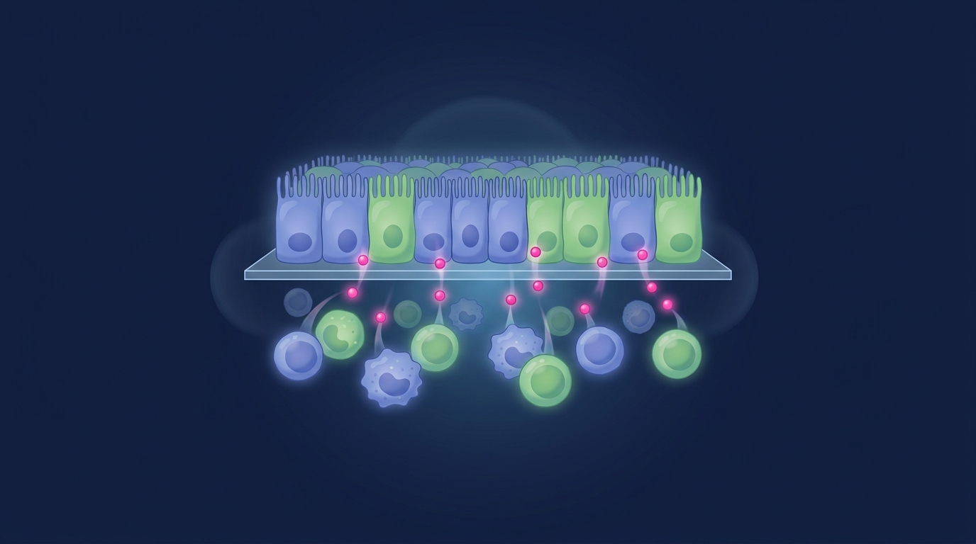
Epithelium on the transwell membrane, THP-1 macrophages in the basal chamber, and inflammatory cytokines passing between the two compartments.

The immune cells drive the barrier breakdown
Under LPS and IFN-gamma, the epithelial barrier holds in monoculture but collapses when THP-1 macrophages are present. Transepithelial electrical resistance, or TEER, drops sharply, alongside reduced viability and a surge of inflammatory cytokines.
That contrast confirms the damage is immune driven, exactly the mechanism at the heart of inflammatory bowel disease.
Corrected TEER, healthy versus inflamed, monoculture versus co-culture.
The barrier damage is preventable with clinical drugs
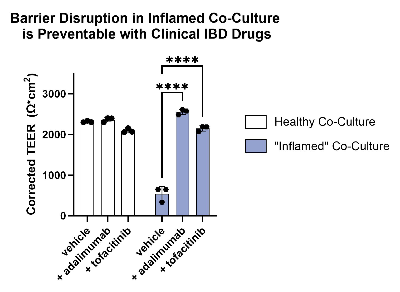
Co-treatment with two FDA approved IBD therapeutics rescues the barrier. Adalimumab, an anti TNF-alpha antibody, and tofacitinib, a JAK inhibitor, both restore TEER in the inflamed co-culture toward healthy levels.
The model responds to drugs that already work in patients. That gives you a trusted human readout for screening the next generation of anti inflammatory therapeutics.
See IBD drug discovery workIn a 2025 study in SLAS Discovery, LPS and IFN-gamma stimulated macrophages compromised the epithelial barrier and raised TNF-alpha, IL-6, and IL-1 beta. Adalimumab and tofacitinib both mitigated the damage and preserved barrier function. Peddibhotla et al., 2025. The model has also supported partner led, peer reviewed IBD drug programs, including published work using RepliGut® co-culture to show a candidate antibody preserved barrier integrity against TL1A driven inflammation.
One gut immune co-culture model, many questions
The same immune and epithelial system supports inflammation, disease, and drug screening work across programs. Pick the application your program needs.
Anti-inflammatory drug screening
Rank therapeutics by how well they protect the barrier against immune driven damage.
IBD disease modeling
Recreate healthy and inflamed colon states to study inflammatory bowel disease biology.
Epithelial and immune interactions
Dissect how macrophage activation and cytokines shape epithelial injury and repair.
Immune relevant biomarkers
Profile cytokine release and barrier endpoints to surface mechanisms and markers.
Barrier integrity readouts
Track TEER and viability as the epithelium responds to compounds and stressors.
Mechanism of injury studies
Test where a compound acts along the immune to epithelial damage pathway.
Why the model holds up
Human relevance
Primary human colon epithelium and human macrophages, not an animal surrogate.
Quantitative endpoints
TEER, viability, and cytokine release give hard numbers in every well.
Reproducible and high throughput
A 96-well format with lot to lot consistency, validated against approved drugs.
Human relevant models also match where regulators are heading. The FDA now actively advances New Approach Methodologies for preclinical testing.
Run it in your lab, or have us run it
RepliGut® Co-Culture kit
The CCT-96W kit adds co-culture medium and a transwell receiver plate to the RepliGut® Planar Transverse Colon model. You run the assay in your own lab, and THP-1 cells are sourced separately.
Explore kitsFull-service screening
Have our scientists run the inflammation and drug screening study for you and deliver TEER, viability, and cytokine data. See our InflammaScreen service.
Explore servicesBuilt on RepliGut® Planar. Studying epithelial renewal and repair instead? See RepliGut® Crypt, or browse the full applications hub.
Screen anti-inflammatory drugs on human gut tissue
Tell us your compound and your question. We will help you design the right RepliGut® Immune Co-Culture study.
Inquire now